Associate Professor and Director of Biomedical Imaging

The hippocampus is a brain region that is critically involved in allowing us to remember places and events. It is damaged early in the biological course of Alzheimer’s disease in a stereotypical spatial pattern: first one specific part of the structure is damaged, then a second specific part, and so on. For this reason, I have worked on ways to quantify localized damage to individual hippocampus parts.
My graduate student Jing Xie further developed a method called Localized Components Analysis (LoCA) that was originally developed by myself, Dan Alcantara, and Nina Amenta of UC Davis Computer Science. LoCA provides a set of measurements describing how much individual parts of the hippocampus is damaged compared to a population average hippocampus. Here is an example in which one particular hippocampus part, shown in blue, is assigned such a measurement (A, L, M, P = anterior, lateral, medial, and posterior directions in the head). When the measurement takes on small values (left hand side of the figure) that part of the structure is relatively small, and when the measurement takes on large values that part of the structure is relatively large (right hand side of the figure).

We showed that these hippocampus part measurements are related to cognitive function in an intuitive way: the measurements covering parts of the hippocampus damaged earliest by Alzheimer’s disease, are associated with the aspects of cognitive function affected earliest by Alzheimer’s disease. In addition, we showed that these local measurements are more strongly associated with the Alzheimer’s disease pathology in the brain than the total volume of the hippocampus is. These results suggest that localized hippocampus measurements might play a useful role in the diagnosis of Alzheimer’s disease as well as clinical trials of disease-modifying therapies.
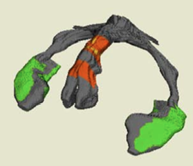
I also worked with Dong Young Lee, a visiting faculty member of Charlie DeCarli’s lab, to show that localized hippocampus measures can lead to new insights about the biological course of Alzheimer’s disease. He showed that localized damage to a part of the hippocampus damaged early in the course of Alzheimer’s disease (shown in green below on the left and right hippocampi) is associated with degeneration of an axon tract (the fornix, shown in red below) through which information flows out of that area to the rest of the brain. While most Alzheimer’s disease research focuses on damage to neurons such as those in the hippocampus, this finding emphasized that breakdown of axons-- the “communication wires” of the brain—plays a prominent role in the disease as well.

Publications:
J. Xie, D. A. Alcantara, N. Amenta, E. Fletcher, O. Martinez, M. Persianinova, C. DeCarli, and O. T. Carmichael. Spatially-Localized Hippocampal Shape Analysis in Late-Life Cognitive Decline. Hippocampus 2009 Jun;19(6):526-32.DOI Also appears in Proceedings MICCAI Workshop on Computational Anatomy and Physiology of the Hippocampus (CAPH), September 6, 2008.
Jing Xie, Owen T. Carmichael. Brain Shape Regression Components. Proceedings of the International Conference of the IEEE Engineering in Medicine and Biology Society (EMBC 2012). DOI
O Carmichael, Jing Xie, M.S.; Evan Fletcher, PhD; Baljeet Singh; Charles DeCarli; Alzheimer’s Disease Neuroimaging Initiative. Localized hippocampus measures are associated with Alzheimer pathology and cognition independent of total hippocampal volume. Neurobiology of Aging, 2012 Volume 33, Issue 6, Pages 1124.e31-1124.e41. DOI
D. Alcantara, O. T. Carmichael, W. Harcourt-Smith, K. Sterner, S. Frost, R. Dutton, P. Thompson, E. Delson, N. Amenta. Exploration of Shape Variation Using Localized Components Analysis. IEEE Transactions on Pattern Analysis and Machine Intelligence (PAMI) 2009 Aug;31(8):1510-6. DOI
Dong Young Lee, Evan Fletcher, Owen Carmichael, Baljeet Singh, Dan Mungas, Bruce Reed, Oliver Martinez, Michael Buonocore, Maria Persianinova, Charles DeCarli. Sub-regional hippocampal injury is associated with fornix degeneration in Alzheimer’s disease. Front Aging Neurosci. 2012;4:1. DOI